

Ranking Osteopathic Lesions Using the LMO Inhibitory Balance Test : Neurophysiological Hypotheses and Clinical Interpretation

Concept Published on Oct 26, 2026

During an osteopathic examination, a patient often presents with several lesions (somatic dysfunctions) distributed across different regions of the body. The practitioner's question is therefore not simply “Where are the lesions?” but, above all, “Which should be treated first?” Identifying a “primary lesion”, however, requires an explicit method of comparison and ranking. The Lien Mécanique Ostéopathique (LMO), known in English as the Mechanical Link and developed by Paul Chauffour from the 1970s onwards[1], provides a practical tool for this purpose: the inhibitory balance test.


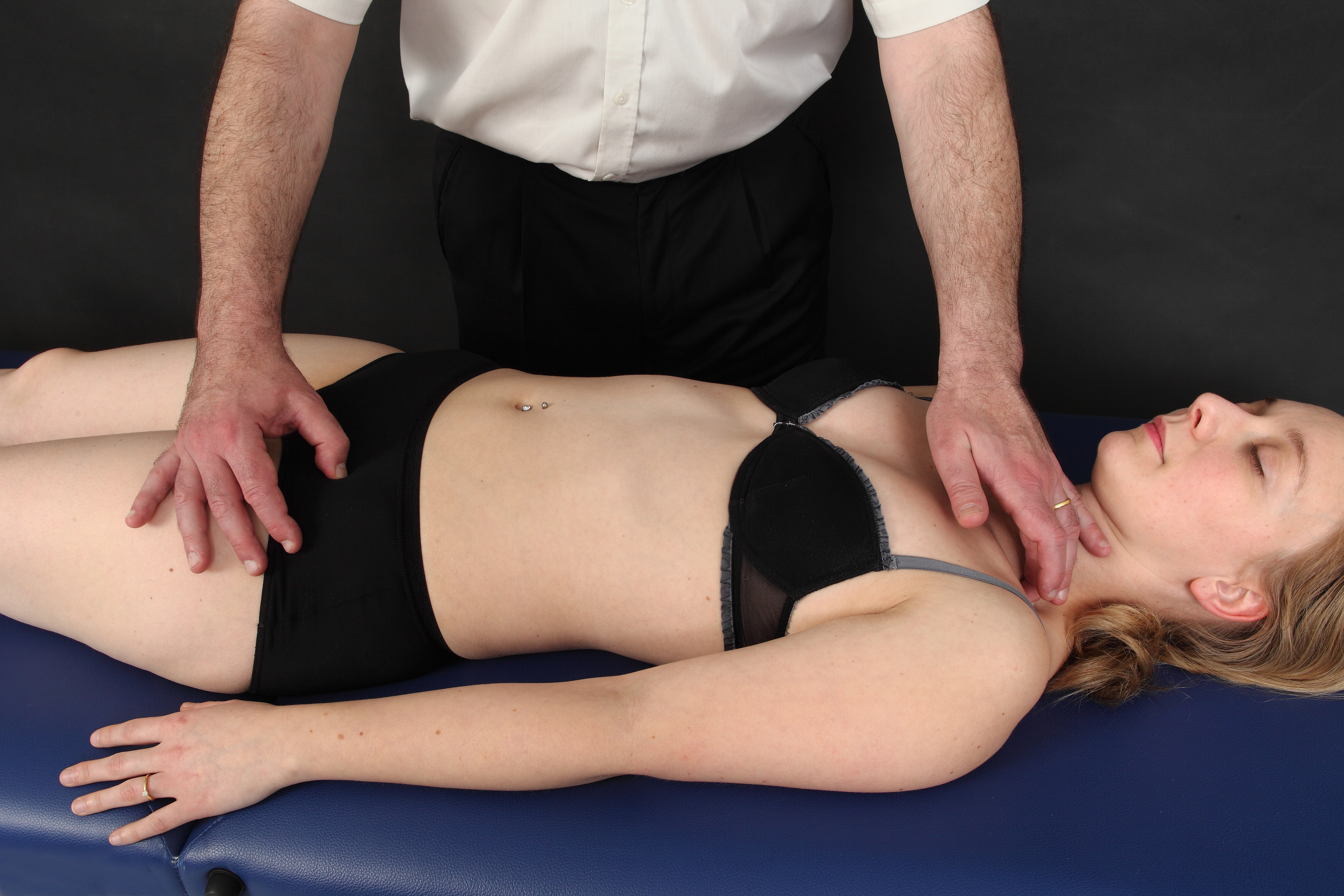
This test compares two osteopathic lesions by applying gentle tension to both simultaneously. The practitioner then perceives a decrease in resistance in one, while resistance in the other persists or appears to increase. By repeating these comparisons, LMO ranks the patient's lesions until the “dominant of the dominants”[2] is isolated. This is termed the primary lesion[3] and is the lesion with which treatment should begin.


This article examines the neurophysiological mechanisms that may contribute to this palpatory phenomenon. It draws particularly on the work of Bicalho et al.[4], who formalised mechanistic hypotheses for inhibitory tests in osteopathy, as well as on classical models of somatic dysfunction (Korr, Van Buskirk and Fryer) and the dominant principle proposed by the Russian physiologist Alexey Ukhtomsky, often cited as a theoretical framework for this phenomenon.


[1]Chauffour P, Guillot JM. Le Lien Mécanique Ostéopathique, substrat anatomique de l’homéostasie. Paris, Maloine, 1985 ; Chauffour P, Prat E. Mechanical Link: Fundamental Principles, Theory and Practice in Osteopathy. Berkeley, North Atlantic Books, 2002.

[2] The dominant lesion is the main restriction within a functional unit.

[3] The highest degree of tissue resistance among the patient's osteopathic lesions on the day of the consultation.

[4]Bicalho E, Vieira L, Makita DK, Rivas L. « Inhibitory Tests as Assessment Tools for Somatic Dysfunctions: Mechanisms and Practical Applications ». Cureus, 2020, 12(4):e7700.
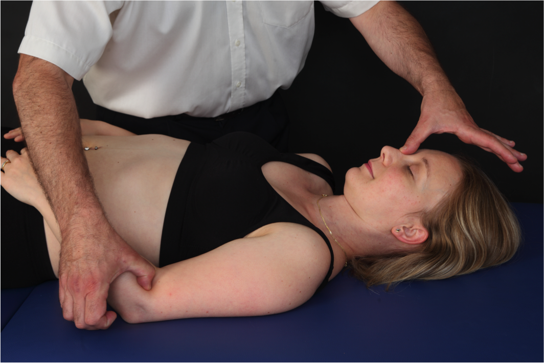
 

The inhibitory balance test: simultaneous tension applied to two lesions and a differential response


A Three-Level Framework

To avoid confusing physiological plausibility with evidence of a mechanism, this article distinguishes three levels of discussion.


1.   Clinical level — what is observed. During the inhibitory balance test, resistance perceived beneath the practitioner's hand decreases at one lesion and persists, or appears to increase, at the other. This palpatory phenomenon is consistently reported by practitioners trained in the method.


2.   Physiological level — what is plausible. Simultaneous manual stimulation of two areas activates cutaneous, proprioceptive, interoceptive and fascial afferents that may modulate spinal excitability, muscle tone, pain and the perception of threat. These mechanisms, described by Bialosky et al. for manual therapy in general and proposed by Bicalho et al. for inhibitory tests, remain hypotheses consistent with current evidence.


3.   Interpretative level — what belongs to clinical interpretation. Within LMO, the lesion whose resistance persists or appears to increase is interpreted as “dominant” or “primary”. This ranking guides the order of treatment.


What Is Observed

1 . The Osteopathic Lesion Revisited


Somatic dysfunction — or osteopathic lesion, in historical terminology — is classically defined by the TART criteria: Tissue texture change, Asymmetry, Restriction of motion and Tenderness. It is described as a connective tissue alteration that develops through an inflammatory process (acute phase) followed by fibrosis (chronic phase), leaving a palpable tissue imprint.


Two criteria allow a lesion to be identified by palpation: a loss of tissue elasticity and increased neurological reactivity, perceived as a defensive reflex response and heightened mechanosensitivity. An “active” lesion, characterised by heightened neurological vigilance and a distinct barrier, is therefore distinguished from a “passive”, adaptive or compensatory lesion, in which tissue resistance is present but the reflex component is attenuated. It is this active, reflex component — rather than the tissue's mechanical density alone — that the inhibitory balance test explores.


2 . From the Tension Test to the Inhibitory Balance Test


LMO diagnosis relies on two complementary techniques. The tension test gently and precisely loads an individual structure to assess its elasticity: normal suppleness, moderate resistance (passive lesion) or a distinct barrier (active lesion). It therefore provides information about both a physical parameter (tissue elasticity) and a reflex parameter (reactivity).

The inhibitory balance test is the second step: it compares two previously identified lesions by applying tension to both simultaneously, using a lighter and subtler contact than in the tension test. One lesion releases beneath the hand, while the other resists or even becomes more pronounced. This ranking can proceed in two stages: first, comparing lesions within a single functional unit (spine, peripheral structures, viscera, cranium, etc.) to identify its dominant lesion; then comparing the dominant lesions of each functional unit to identify the primary lesion, the “dominant of the dominants”[1].


[1] Méchanical Link. “Diagnosis and treatment of the osteopathic lesion using the LMO approach". lmosteo.com, 2021.


Inhibitory balance test between the radial head and the nasal bone.


What Is Physiologically Plausible

To understand how two distant lesions may “communicate” beneath the practitioner's fingers, we must return to the segmental (metameric) organisation of the nervous system, established during embryogenesis.


1. Metameric Organisation as an Available Anatomical Substrate


Somitogenesis organises the paraxial mesoderm into somites on either side of the neural tube. These differentiate into three structures: dermatomes (dorsal dermis), sclerotomes (part of the axial skeleton) and myotomes (skeletal muscles of the trunk and limbs). Fasciae derive from embryonic mesenchyme, originating mainly from the paraxial or lateral mesoderm depending on the region, with a neural crest contribution in the craniofacial region and part of the neck[1]. This organisation contributes to the segmental arrangement of somatic structures and their innervation. In parallel, the viscera connect to several spinal cord levels, particularly through sympathetic nervous system pathways.


Functionally, a single spinal cord segment receives sensory afferents from different tissues: skeleton, muscles, viscera, blood vessels and skin. Somatic and visceral structures can therefore share spinal integration circuits without having the same embryological origin. Here, the metameric link refers to the sharing of one or more spinal connection levels rather than strict membership of a single anatomical territory. This convergence of somatic and visceral afferents provides a substrate for referred pain and interactions between these sensory inputs. Within LMO, it supports the hypothesis that an osteopathic lesion may, through spinal circuits, influence the perceived reactivity of another tissue sharing some of its connection levels. When the two areas tested share no spinal cord level — for example, the nasal bone and the radial head — segmental convergence alone is insufficient: the perceived interaction would then require supraspinal integration.


2.  Central Sensitisation and the Facilitated Segment


In 1975, Irvin Korr proposed the “facilitated segment” model[1]: repeated afferent bombardment from proprioceptors in a dysfunctional area would lower the “synaptic resistance” of the corresponding spinal cord segment and facilitate impulse transmission to effectors (muscles and viscera). Van Buskirk expanded this model in 1990[2], emphasising the role of nociceptors and neurogenic inflammation following mechanical, chemical or thermal stress.


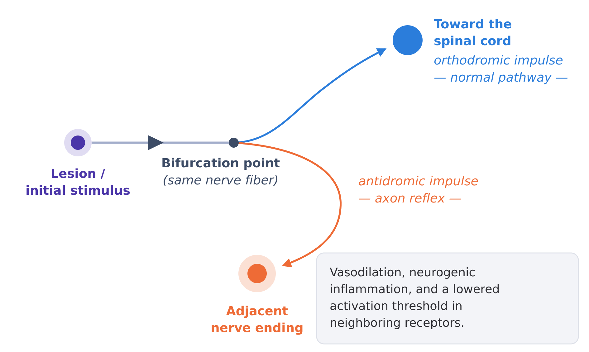
Pain physiology has since refined this mechanism through the concept of central sensitisation[3]. Wide dynamic range (WDR) neurons in the dorsal horn receive convergent cutaneous, deep somatic and visceral afferents from the same segment. When tissue is injured or inflamed, antidromic activity[4], termed the axon reflex[5], sustains local neurogenic inflammation and lowers the activation threshold of peripheral receptors (peripheral sensitisation), producing hyperalgesia or even allodynia in the affected area. These two effects are not contradictory but sequential: the inflammation itself need not be painful at the moment it appears; rather, the reduction in threshold makes subsequent stimulation more painful than it would normally be. After sunburn, the skin becomes red and warm — this is local inflammation — but it is not the redness itself that hurts. Rather, lukewarm shower water or the simple touch of clothing, normally painless, becomes painful the following day because inflammation has lowered the activation threshold of the surrounding cutaneous receptors: this is allodynia.

[1] Fasciae and the muscles they envelop do not necessarily share the same embryological origin.

[2]Korr IM. « Proprioceptors and somatic dysfunction ». Journal of the American Osteopathic Association, 1975, 74:638-650.

[3]Van Buskirk RL. « Nociceptive reflexes and the somatic dysfunction ». Journal of the American Osteopathic Association, 1990, 90:792-809.

[4]Woolf CJ. « Central sensitization: Implications for the diagnosis and treatment of pain ». Pain, 2011, 152(3 Suppl):S2-S15.

[5] An antidromic impulse travels in the opposite direction to the usual pathway. In the axon reflex, it travels from a sensory fibre branch point towards neighbouring peripheral terminals.

[6]Howell JN, Willard F. « Nociception: new understandings and their possible relation to somatic dysfunction and its treatment ». Ohio Research and Clinical Review, 2005, 15:35.


The axon reflex. At the fibre branch point, antidromic activity sustains local neurogenic inflammation.


This local mechanism is accompanied by a central mechanism, the dorsal root reflex: when the spinal cord is sensitised, it can itself send impulses back towards the periphery. A spinal cord segment “on alert” can thus sustain inflammation in the various tissues it innervates, rather than only at the original lesion. This mechanism remains limited, however, to tissues within the same segment.


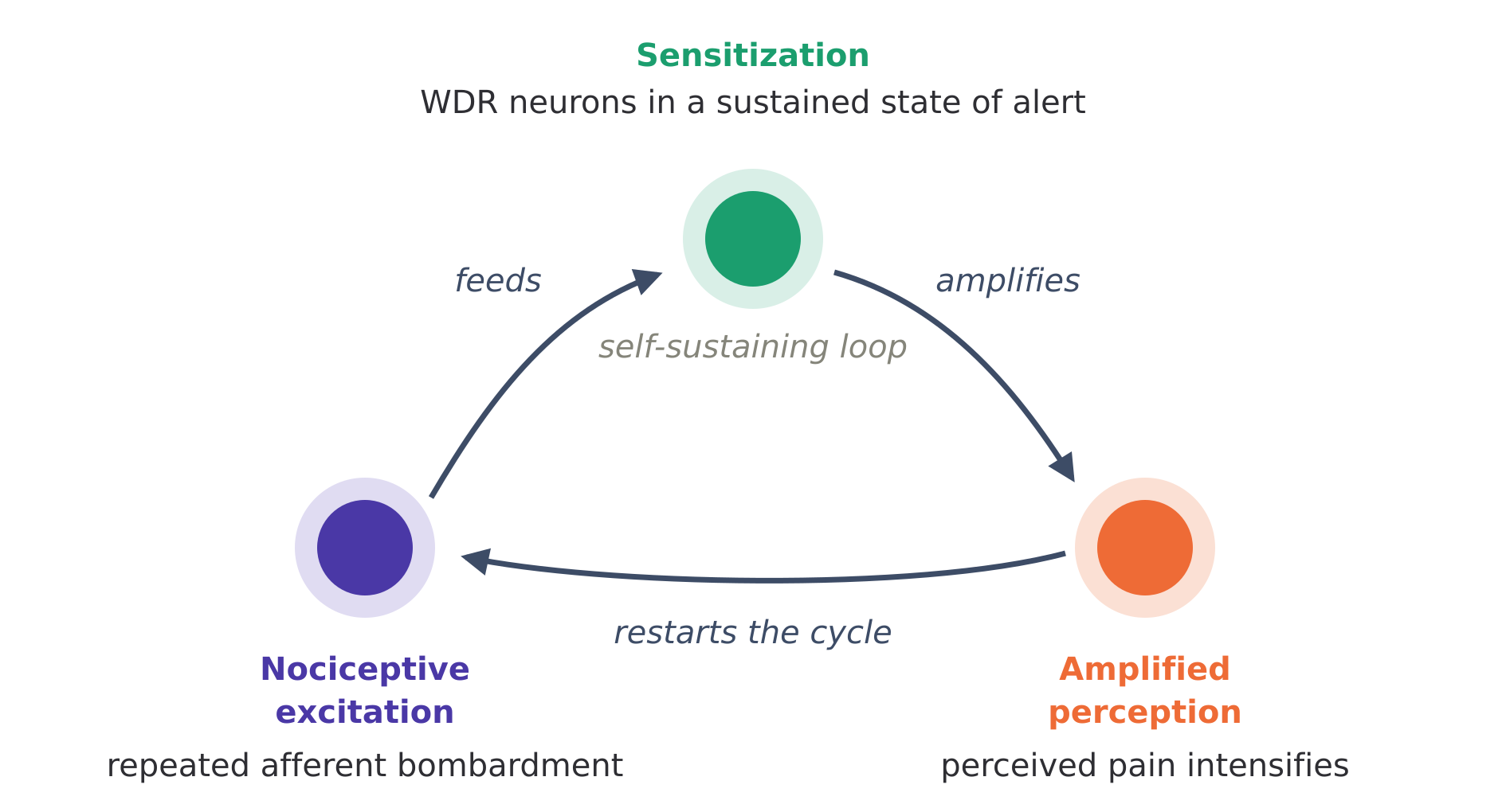
If this nociceptive bombardment is sufficiently intense or prolonged, it can keep the WDR neurons themselves in a sustained state of alert: this is central sensitisation, which some authors describe as “spinal segmental sensitisation”. This self-sustaining cycle forms a kind of “pain reflex loop”: nociceptive excitation sustains sensitisation, which in turn amplifies the perception of that same excitation.


The pain reflex loop: how nociceptive excitation and sensitisation sustain each other.


This theoretical framework sheds light on the inhibitory balance test: certain osteopathic lesions, particularly when active or painful, may be considered not as strictly local phenomena but as the possible peripheral expression of a neural network capable of sustaining hyperexcitability. It is this “network” dimension that the test explores by applying tension to two areas simultaneously.


3. Bialosky's Model from Mechanical Force to Neurophysiological Response


Beyond the specifically osteopathic field, the model proposed by Bialosky et al.[1] for manual therapies as a whole suggests that a mechanical force applied by the therapist triggers a cascade of peripheral, spinal and supraspinal neurophysiological responses that may influence clinical parameters such as pain, muscle tone or mobility. This model also incorporates non-specific factors — patient expectations, the therapeutic relationship and the care context — whose influence on observed neurophysiological responses is now well established.


This framework calls for caution: what a practitioner perceives during a manual test may result from a combination of mechanical, neurophysiological and contextual mechanisms that are difficult to isolate in clinical practice. A local mechanical stimulus, such as that used in the inhibitory balance test, is never confined to a single level of the nervous system or to the structure directly stimulated.


4. Possible Mechanisms of Touch-Evoked Inhibitory Responses


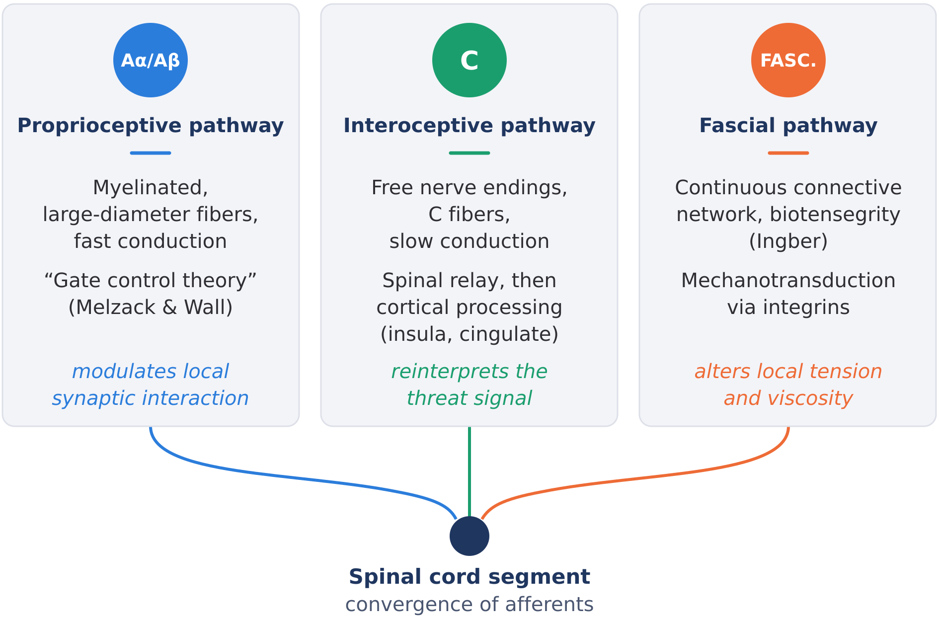
In their 2020 review, Bicalho et al.[1] propose three non-exclusive and probably interacting mechanisms to explain the reflex responses observed during manual inhibitory tests in osteopathy


[1]Bialosky JE, Bishop MD, Price DD, Robinson ME, George SZ. « The mechanisms of manual therapy in the treatment of musculoskeletal pain: a comprehensive model ». Manual Therapy, 2009, 14(5):531-538 ; actualisé par Bialosky JE et al. « Unraveling the Mechanisms of Manual Therapy: Modeling an Approach ». Journal of Orthopaedic & Sports Physical Therapy, 2018, 48(1):8-18.

[2]Bicalho et al., op. cit., note 4.


Three plausible touch-evoked pathways

based on the model proposed by Bicalho et al. (2020)

    4.1 The Proprioceptive Pathway and Gate Control Theory


Light manual contact activates low-threshold tactile and proprioceptive mechanoreceptors whose signals travel along large-diameter myelinated fibres (types I and II, or Aα and Aβ) at high conduction velocities (30–120 m/s, depending on fibre type). Their collaterals activate interneurons in the dorsal horn, where the C fibres involved in sensitisation converge. Because proprioceptive input arrives faster and more intensely than nociceptive input, it transiently alters synaptic interactions and may reduce neural sensitisation in the segment concerned. This is the mechanism described by Melzack and Wall's gate control theory[1], which remains a key conceptual framework in pain physiology.


    4.2 The Interoceptive Pathway and Free Nerve Endings


A second receptor network, the free nerve endings (approximately 80% of sensory receptors in fascial tissue)[1], is connected to unmyelinated C fibres, which conduct slowly (approximately 1 m/s) but have a strong capacity for sensitisation. These multimodal receptors respond to mechanical, thermal, chemical and nociceptive stimuli. Slow, light and non-aggressive touch may alter the interpretation of their message: a potentially threatening signal becomes a “pleasant”, non-nociceptive signal. This stimulation activates cortical areas such as the insular cortex, anterior cingulate cortex and prefrontal cortex, while modulating amygdala activity. These regions contribute to the interpretation of interoception, emotions and pain: their activation may alter the initial conditions of what Ronald Melzack called the “neuromatrix”[2], promoting a reinterpretation of the pain signal and contributing to central desensitisation.


    4.3 Local and Global Changes in Fascial Tissue


Fascia forms a continuous connective tissue network linking all the body's structures, from the deepest to the most superficial[1]. Its viscoelastic properties allow it to adapt continuously to mechanical stresses, both locally and globally: this is the principle of biotensegrity. Forces applied by the hand are transmitted from the extracellular matrix into cells through integrins (mechanotransduction)[2], which can modulate cell growth, differentiation and gene expression[3]. A brief mechanical stimulus applied to a dysfunctional area may therefore transiently alter connective tissue densification, fluid dynamics and the activation threshold of mechanosensitive receptors within the extracellular matrix, with local effects (vasodilation and changes in viscosity) and distant effects through the continuity of the fascial network.

These three pathways — proprioceptive, interoceptive and fascial — are likely to act together and almost instantaneously as soon as the practitioner places their fingers on a lesioned area. They transiently modify the afferent traffic converging on the spinal cord, explaining the rapid response perceived during the inhibitory balance test.


[1]Melzack R, Wall PD. « Pain mechanisms: a new theory ». Science, 1965, 150(3699):971-979 ; voir aussi Mendell LM. « Constructing and deconstructing the gate theory of pain ». Pain, 2014, 155:210-216.

[2]Schleip R. « Fascial plasticity – a new neurobiological explanation: part 1 ». Journal of Bodywork and Movement Therapies, 2003, 7:11-19.

[3]Melzack R. « Pain and the Neuromatrix in the Brain ». Journal of Dental Education, 2001, 65(12) :1378-1382.

[4]Guimberteau JC, Armstrong C. Architecture of Human Living Fascia: Cells and Extracellular Matrix as Revealed by Endoscopy. Edinburgh, Handspring Publishing, 2015.

[5]Ingber DE. « Tensegrity-based mechanosensing from macro to micro ». Progress in Biophysics and Molecular Biology, 2008, 97:163-179.

[6] These effects, however, unfold over hours or days, rather than within the few seconds of the test.


What Belongs to Osteopathic Interpretation

1. Alexey Ukhtomsky's Dominant Principle as a Historical Theoretical Framework and Functional Analogy for LMO


In 1923, the Russian physiologist Alexey Ukhtomsky (1875–1942) formulated the concept of the “dominant”[1], based on a chance laboratory observation made as early as 1904: in a dog in the process of defecating, electrical stimulation of the motor cortex no longer produced the usual limb movements but instead intensified the ongoing act of defecation. From this, he derived a general principle of nervous system function.


[1] Ukhtomsky A. Dominanta [The Dominant]. St Petersburg, Piter, 2002 (reprint; original text published in 1923).

“The dominant is a constellation of nerve centres with heightened sensitivity to excitation, located at different levels of the brain and spinal cord, as well as in the autonomic nervous system.” — Alexey Ukhtomsky

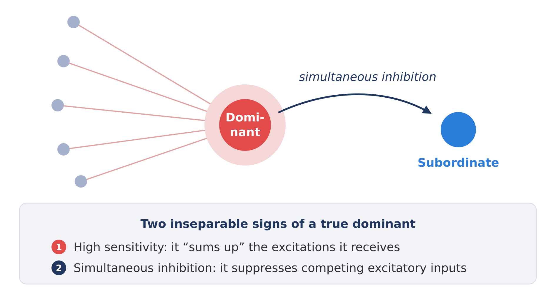
For Ukhtomsky, a true dominant always combines two inseparable characteristics: heightened sensitivity arising from the accumulation and persistence of stimuli (it “sums excitations” rather than responding solely to their intensity), and simultaneous inhibition of competing sources of excitation. Without both signs, he states, one cannot speak of a true dominant.

Ukhtomsky’s dominant principle

Two neural networks competing for the same excitations


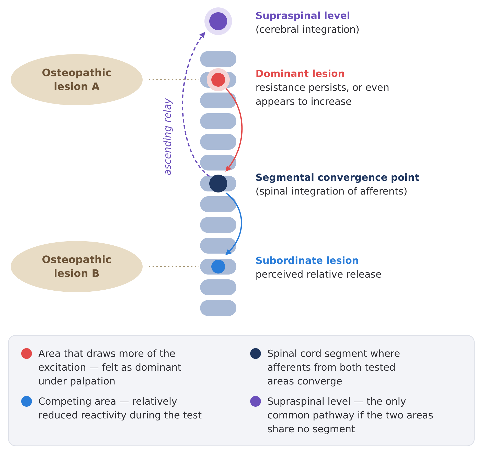
The parallel with the inhibitory balance test is striking: when tension is applied simultaneously, the tension perceived at the dominant lesion persists or appears to increase, while that of the subordinate lesion decreases — a pattern reminiscent of the paired features described by Ukhtomsky[1]. Each osteopathic lesion can therefore be considered a physiological dominant in relation to the surrounding healthy tissues or to other lesions. This concept, which historically predates Korr's facilitated segment, converges with more recent models of central sensitisation: hyperalgesia and allodynia can be interpreted as the somatic expression of a dominant in Ukhtomsky's sense[2].


More recent imaging data point in the same direction: functional MRI studies have shown changes in cerebral perfusion and brain correlates of interoception after osteopathic treatment[3]. They suggest that manual correction changes not only local tissue elasticity but also the functioning of central neural networks — what Melzack termed the neuromatrix[4].


2. From Neurophysiology to Manual Perception During the Test


Bringing these explanatory levels together, the following physiological sequence can be described. Simultaneous tension applied to two lesioned areas generates an influx of mechanoreceptive, proprioceptive and interoceptive information in each. Integrated at spinal, supraspinal and autonomic levels, these afferents may transiently alter tone, the perception of threat and perceived tissue resistance.


One may then hypothesise that the area associated with the most active, or most strongly organised, sensitisation network retains more pronounced resistance: beneath the fingers, tension persists or even increases. Conversely, reactivity in the competing area transiently decreases, creating an impression of release.

The test therefore does not directly measure neurological causality between the two lesions. It reveals a differential resistance response between two areas subjected to simultaneous manual loading.


This phenomenon explains why experienced practitioners emphasise the need for extremely light contact[1]: the inhibitory balance test does not compare the mechanical properties of two tissues, such as density or elasticity (bone does not have the same density as a viscus), but rather the relative neurophysiological reactivity of two restricted areas. Within LMO's methodological and teaching framework, by successively comparing the diagnosed lesions and then the lesions identified as dominant in each body region, the practitioner converges on a single “dominant of the dominants”, termed the primary lesion. This presumably reflects the somatic expression of the most active neurophysiological network in that patient at that particular moment.


[1]Ukhtomsky A. Dominanta [La dominante]. Saint-Pétersbourg : Piter ; 2002, p. 199 cité par Bodykhov M. « Le principe de dominante d'Alexey Ukhtomsky ». lmosteo.com, 2023.

[2]Fryer G. « Somatic dysfunction: an osteopathic conundrum ». International Journal of Osteopathic Medicine, 2016, 22:52-63.

[3]Tamburella F et al. « Cerebral Perfusion Changes After Osteopathic Manipulative Treatment: A Randomized Manual Placebo-Controlled Trial ». Frontiers in Physiology, 2019, 10 :403 ; Cerritelli F et al. « Effect of Manual Approaches with Osteopathic Modality on Brain Correlates of Interoception: an fMRI Study ». Scientific Reports, 2020, 10 :3214.

[4]Melzack, op. cit., note 16.

[5]Dohata M. « Force d'appui optimale pour le test de balance inhibitrice ». lmosteo.com, 2023.


Clinical Implications and Methodological Limitations

This three-level reading sheds light on a clinical observation repeatedly reported by LMO practitioners: treating the primary lesion frequently leads to the spontaneous normalisation of numerous distant secondary or adaptive lesions without treating them directly. If a substantial proportion of these secondary lesions results from central sensitisation sustained by the dominant network, then mechanical and neurological correction of that network — for example, using the recoil technique (a brief, low-amplitude, high-velocity impulse applied to a restriction that has first been placed under tension) — may reduce sensitisation of WDR neurons in the spinal cord segments concerned and thus “switch off” the peripheral manifestations dependent on them.


A critical stance must nevertheless be maintained. Bicalho and colleagues[1] themselves emphasise that these mechanisms remain plausible hypotheses constructed by analogy with general neurophysiology, rather than direct demonstrations of how the inhibitory balance test itself works. Reliability studies exist for the LMO tension test[2], but specific scientific validation of the inhibitory balance test — inter-practitioner reproducibility, sensitivity and specificity — remains an open area of research.


[1]Bicalho et al., op. cit., note 4.

[2]Hafen-Bardella C. Reliabilitätsstudie über die Befunderhebung der Wirbelsäule nach der Methode der Lien Mécanique Ostéopathique. Master Thesis, Donau-Universität Krems, 2009 ; Hafen G. Reliabilitätsstudie über die Befunderhebung der Knochen und Gelenke der Extremitäten nach der Methode des Mechanical Link. 2018 ; Kühn L. Follow-up-Studie über die Reliabilität der Befunderhebung der Wirbelsäule nach der Methode des Mechanical Link. 2018.


Conclusion

Although the inhibitory balance test involves a technically very simple manoeuvre, it engages a coherent set of neurophysiological mechanisms: metameric organisation originating in somitogenesis, central sensitisation and the facilitated segment, the proprioceptive, interoceptive and fascial pathways of therapeutic touch, and Ukhtomsky's dominant principle. Together, these elements help explain why the simultaneous comparison of two osteopathic lesions produces a palpatory phenomenon regularly described by LMO practitioners: persistence, or even perceived intensification, of the dominant lesion and release of the subordinate lesion.


The resulting ranking therefore belongs to clinical interpretation: it guides the order of treatment but does not demonstrate neurophysiological causality between the lesions. The test probably does not measure “the primary lesion” as such, but rather a differential resistance response between two areas subjected to simultaneous manual loading. It is this response, interpreted within the LMO framework, that leads the practitioner to designate a restriction as dominant or a treatment priority.


The teaching value of the inhibitory balance test thus lies in explicitly relating three levels: clinical observation, physiological plausibility and osteopathic interpretation. Distinguishing these levels, rather than conflating them, makes it possible to offer an account that is rigorous, critical and usable in practice.


Olivier Samson DO


This article was written with the support of artificial intelligence tools to critically examine, structure and improve reasoning initially developed by the author, as well as for literature searches and the creation of some illustrations.

The content was subsequently reviewed, validated and expanded by the author.

Linked articles
MECHANICAL LINK : FOUNDATIONS
Mar 28, 2021
Mechanical Link (in French, Lien Mécanique Ostéopathique or LMO) was ...
READ MORE
MECHANICAL LINK : EVOLUTION AND PERSPECTIVES
Apr 1, 2021
The last twenty years have largely been devoted to the publication of d ...
READ MORE
TEACHING LMO
Jun 13, 2021
Mechanical Link (LMO) is a practical science that must be learned prope ...
READ MORE
DIAGNOSIS AND TREATMENT OF THE OSTEOPATHIC LESION ACCORDING TO THE LMO APPROACH
Oct 30, 2021
ALCHEMY WITH THREE INGREDIENTS ...
READ MORE
Osteopathic regulation of the body’s systems using the Extraordinary Vessels Part 1: traditional concept
Jun 24, 2024
In osteopathy, we pay a lot of attention to the 3 anatomical links betw ...
READ MORE
Osteopathic regulation of major body systems through Extraordinary Vessels Part 2: anatomical and physiological reading
Sep 30, 2024
Trying to find connections between Extraordinary Vessels and academic p ...
READ MORE
Osteopathic regulation of major body systems through Extraordinary Vessels Part 3: clinical study Sympathetic system
Oct 31, 2024
From an osteopathic point of view, each Extraordinary Vessel could be d ...
READ MORE
Osteopathic regulation of major body systems through Extraordinary Vessels Part 4: clinical study Parasympathetic system
Dec 6, 2024
Following on from our previous article, let's recall that from an osteo ...
READ MORE
Osteopathic regulation of major body systems through Extraordinary Vessels Part 5: osteopathic regulation protocol
Jan 13, 2025
With Osteopathic Mechanical Link, which could be described as digital o ...
READ MORE